| |
Pediatric cholelithiasis and breastfeeding difficulties: A chiropractic case report
By Michelle A. Hubbard, MChiro
Michelle A. Hubbard, MChiro, private practice, Camden, NSW, Australia
Contact: hubbardmichelle@hotmail.com
Abstract: Through presenting the case of a 7-week-old the object of this report is (1) to create awareness of the increasing rate of cholelithiasis in the pediatric population, and (2) to outline how chiropractic care assisted in the resolution of breastfeeding difficulties. Design: A case report. Clinical Features: Following a chiropractic health history and physical examination it was identified that the breastfeeding difficulties were a consequence of a combination of issues. The infant was found to have limited left rotation of the neck, a result of upper cervical subluxations and cranial misalignments, and ankyloglossia (tongue-tie). Further investigation via stool analysis, abdominal ultrasound and blood testing led to the diagnosis of fetal cholelithiasis with an underactive gallbladder. Intervention & outcomes: Chiropractic adjustments were implemented to correct the cervical and cranial motion. Mother and child were also placed on daily probiotic supplementation. After one month of weekly chiropractic care the child was found to have normal cervical range of movement. A frenotomy was performed at 10 weeks old. The infant displayed complete resolution of the breastfeeding difficulties. At 11 weeks the infant was placed on 1.2ml Ursofalk ursodeoxycholic acid (bile acid) twice a day and 0.2ml colecalciferol (VitD) daily. This continued for one month. No further treatment was implemented for the cholelithiasis. Conclusion: As primary care practitioners it is essential that chiropractors recognize and understand the pathophysiology of gallbladder disease in the pediatric population. It is possible that the presenting symptoms may be misdiagnosed and therefore lead to inappropriate treatment. In this case a multidisciplinary approach was required to manage the various presentations. Chiropractic care resolved the biomechanical component of the breastfeeding difficulties which occurred concurrently with the cholelithiasis.
Key words: chiropractic, pediatrics, breastfeeding, subluxation, fetal, cholelithiasis, gallstones, gallbladder.
Introduction
Cholelithiasis, more commonly known as gallstones, is a common disorder of the digestive system affecting approximately 20% of adults over 40 and 30% aged over 70 years1.
Once known as an adult disease, cholelithiasis has more recently been found to have no age discrimination. The prevalence is on the rise in the pediatric population, an outcome of increased ultrasonography use in this age group and rising childhood obesity levels2. Studies indicate a rate of between 0.13%3 and 1.9%4.
Often, a chiropractor is the initial health practitioner to examine a child in pain. In a neonate or infant who cannot describe the location or type of pain they are experiencing, the presentation of symptomatic cholelithiasis may possibly be misdiagnosed. Colic, reflux, breastfeeding jaundice and food intolerances are all plausible diagnoses for symptoms of cholelithiasis.
It is therefore the purpose of this report to increase awareness of gallbladder disease in the pediatric patient through outlining the clinical presentation of a 7-week-old infant who initially presented to the chiropractic office to address colic and unsettled behavior and was later diagnosed with cholelithiasis and an underactive gallbladder.
This infant was simultaneously experiencing difficulties with breastfeeding. Breast milk contains the perfect composition required for growth, development and immunity5. The world health organization recommends infants be exclusively breastfed for the first 6 months of life, with continued breastfeeding along with suitable complimentary foods for up to 2 years or more6.
Breastfeeding is a synchronized event requiring the infant to suck, swallow and breathe. Six of the 12 cranial nerves; 22 cranial bones (or segments of bones) connecting at 34 sutures; and 60 voluntary and involuntary muscles are used to perform this coordinated activity7. Disruption to the proper function of either the musculoskeletal or nervous systems can therefore impact on an infant’s ability to breastfeed successfully7.
It is therefore imperative that when difficulties with the breastfeeding process arise, they be immediately addressed and corrected to prevent the likely consequence of premature replacement with another food source.
Methods
An online literature search was conducted using the journal databases PubMed, Medline, Science Direct and Index to Chiropractic Literature. Key search words and phrases included ‘cholelithiasis’, ‘gallstones’, ‘pediatric cholelithiasis’, ‘infantile cholelithiasis’, ‘fetal cholelithiasis’, ‘pediatric gallstones’, ‘infantile gallstones’, ‘fetal gallstones’, ‘cholelithiasis + chiropractic’, ‘gallstones + chiropractic’, ’breastfeeding difficulties’, ‘breastfeeding difficulties + chiropractic’. Other internet searches using the above terms were also conducted, as was a manual search of the Journal of Clinical Chiropractic Pediatrics. Articles published between 1985 and 2013 were included.
Case Presentation
A 7-week-old female presented to a chiropractic office to address a 4-day history of what her mother described as tummy pain. Symptoms included green stools that contained mucous, dark yellow urine, being very unsettled while awake, and vomiting after feeds. The vomit was defined as being smelly and sometimes lumpy.
Since the onset of symptoms the infant always had to be held, did not want to sleep on her back, which had previously been the norm, or be in the car seat. Also, the infant was said to regularly bring her knees up to her chest, a practice which had been ongoing since birth.
Prenatal history was unremarkable. Gestation was 39.4 weeks ending in a spontaneous labor. Labor lasted 5 hours and 45 minutes and no intervention was used. Following the birth of a 4.06kg, 54cm long girl, the 29-year-old primiparous mother suffered with retained placenta and post-partum hemorrhage. She was placed on iron supplementation and the antibiotics Metrogyl and GenRx Cephalexin. APGAR scores are unknown.
The baby girl has been breastfed since birth. Her mother describes the infant as sometimes having difficulty suckling, having a preference for the left breast and making a clicking sound as she comes off the right breast. She was feeding every 2 hours with each feed taking 10 minutes. The mother did not suffer from mastitis, cracked nipples or pain while feeding.
Prior to the onset of symptoms, the infant had regular bowel movements that were described as a normal consistency and a mustard color.
Family history includes a maternal aunt having undiagnosed digestive issues and maternal great grandfather having his gall bladder removed during his 50’s.
Clinical Findings
The 7-week-old infant weighed 5.6kg and was 59.5cm long. Temperature was 36.6ºC and mild jaundice of the skin was present.
During the chiropractic appointment the infant was extremely distressed, crying and inconsolable throughout most of the hour long consult. The positive examination findings included a rigid and tender abdomen on palpation, reduced cervical rotation to the left, ankyloglossia, a high palatal arch, an elongated and thickened labial frenulum, dishing of the greater wings of sphenoid (sphenoid extension) and parietal bone overlap. Subluxations present were a right posterior superior occiput (occ-RPS) and right posterior C2 (C2RP). Spasticity of the right suboccipital muscles was also noted.
The initial diagnosis was breastfeeding difficulties as a result of aberrant upper cervical biomechanics (subluxation), cranial misalignment and tongue-tie. The absence of an elevated temperature indicated infection was unlikely therefore the digestive issues were initially thought to be related to a food allergy. However, it was not until further testing was done that an accurate diagnosis was made.
Subsequent to the chiropractic examination, laboratory investigations identified the infant as having an elevated blood bilirubin level. It was following two abdominal ultrasounds that the infant was found to have a gallstone in the neck of the gallbladder. She was diagnosed with fetal cholelithiasis.
At 11 weeks of age, further ultrasounds diagnosed an underactive gallbladder, contracting at just 32% capacity. The medical doctor informed the infant’s mother that the minimum effective level is 35%. At this age, the infant was also identified as having a low Vitamin D level.
Intervention & Outcomes
Chiropractic treatment took place on a weekly basis for 5 weeks then biweekly for 1 session. The objective of the treatment was to restore correct cervical and cranial range of motion which was achieved by the fourth visit. No adverse effects were experienced as a result of the treatment provided.
On the initial assessment the right occiput was found to be subluxated in a posterior and superior direction. Correction was made by applying gentle pressure in anterior and inferior direction for several seconds with the infant supine. This was followed by a gentle rocking motion of the right occiput. C2 was also adjusted this visit with a gentle press and hold on the posterior aspect of the right transverse process. After a few seconds of holding a light thrust in the same direction was applied. A sagittal suture spread was performed by applying light pressure with the fingertips to both sides of the suture in a medial to lateral direction. Lastly, a spheno-basilar flexion mobilization was performed. During this procedure a gloved little finger was placed in the infants’ mouth to apply superior and posterior pressure just posterior to the transverse palatine suture. Pressure was held for 3-5 seconds and repeated 3 times.
As the mother was prescribed antibiotics following labor and the infant was breastfed, both mother and infant were placed on a daily dose of probiotic supplementation. The infant was prescribed 2g once a day of BioCeuticals® BabyBiotic 0+yrs formula and the mother was prescribed one capsule daily of BioCeuticals® UltraBiotic Pregnancy Care.
The day following the first chiropractic treatment, the infant passed one large bowel movement of normal color and consistency. The subsequent bowel movement returned to green.
The same treatment protocol was repeated one week later whereby the infant was found to be having normal bowel movements, was vomiting less and was said to be more settled. During the 3rd and 4th treatment sessions a SOT gallbladder reflex technique was also performed8. By the fourth week the infant was much more settled and cervical range of movement was normal.
At 10 weeks of age a frenotomy was performed to correct the tongue-tie. Following the procedure the infant had a stronger suckle and no longer made a clicking sound coming off the right breast.
The cholelithiasis was monitored from 7 weeks to 4 months of age with the use of abdominal ultrasound (Table 1). It was important to rule out a hemolytic or other specific cause of the cholelithiasis, which is why stool and blood laboratory testing was performed. These tests were negative.
Table 1. Medical management of the infant. |
| Age of Infant |
Medical Intervention |
Outcome |
| 7 weeks |
Stool sample
Blood sample
Two abdominal ultrasounds (one pre and post feeding) |
Negative for infection
Elevated bilirubin. Negative for hemolytic disease.
Gallstone in neck of gall bladder
Pediatrician recommends monitor |
| 9 weeks |
Abdominal ultrasound
Blood sample |
1 Gallstone 2.8x6mm
Results unknown |
| 10 weeks |
Frenotomy |
Suckle improved |
| 11 weeks |
Abdominal ultrasound with dye (cholescintigraphy)
Blood sample |
Gallbladder found to be underactive contracting at 32%
Low vitamin D level. Bilirubin normal.
Infant placed on 0.2ml colecalciferol (VitD) daily and 1.2ml Ursofalk ursodeoxycholic acid (bile acid) twice a day. |
| 4 months |
Abdominal ultrasound
Blood sample |
Stone remains present
Normal. Stopped taking VitD and Ursofalk |
| 5 months |
Hospitalized with fever & vomiting bile |
Gastroenteritis diagnosed. Infant administered Hydralyte™.
Parents identify gall stone in stool. |
Cholescintigraphy is a test done to identify obstruction of the bile ducts and disease of the gallbladder. A radioactive chemical is injected into the body which is handled by the liver like bile9. At 11 weeks old the infant underwent a cholescintigraphy confirming the location of the gallstone and underactivity of the gallbladder. Subsequently the infant was prescribed 1.2ml Ursofalk™ ursodeoxycholic acid twice a day for one month. Ursodeoxycholic acid is a bile acid that is administered in cases of chronic cholestatic liver disease10. Its mechanism of action is to increase bile acid output and bile flow from the liver10, however it is unclear whether this treatment was beneficial. An ultrasound performed at 4 months of age showed the gallstone was still present and the contractility of the gallbladder has not yet been re-tested.
A blood test performed at 11 weeks also identified the infant as having low vitamin D. She was prescribed 0.2ml colecalciferol daily. The next blood reading showed a normal reading so at 4 months the supplementation ceased.
At five months of age the infant was hospitalized with an episode of fever and vomiting bile. A diagnosed of gastroenteritis was made and the infant was placed on a rehydration formula (Hydralyte™). During this period the parents identified what they believed to be a gallstone in the infant’s stool. No follow-up ultrasound has yet been performed to confirm the passing of the stone.
Solid foods were introduced to the infant at 6 months of age to supplement the breastfeeding. Initially the infant experienced vomiting episodes after the intake of solid food. Rice cereal, pear, apple and sweet potato were all found to have the same effect. After two weeks of testing different food the infant was found to accept avocado and pumpkin without vomiting. At 7 ½ months old, the infant was still limited by what she could consume, possibly an indication the gallbladder was still not functioning correctly.
A follow-up chiropractic progress examination was performed when the infant was 6 ½ months. During this examination the infant was found to be happy, symptom free and developing well. She had been rolling since 5 months and was sitting up unsupported for a brief period of time. Cervical range of motion was found to be within normal limits and the infant was having no difficulty breastfeeding. Status of the cholelithiasis and gallbladder contractility remains unknown as no further ultrasounds have been performed.
Discussion
The clinical presentation of pediatric cholelithiasis is varied. Age of the child is one factor that may impact how the disease is expressed, etiology is another. The clinical presentation of the infant described in this paper is unusual for two reasons. Her gallstones were not detected until 7 weeks of age and the symptoms she expressed are uncommon for her age. Fetal cholelithiasis is classically detected in the 3rd trimester via routine obstetric ultrasound and stereotypically remains asymptomatic11,12.
A summary of common clinical presentations at the various ages through childhood is seen in Table 211-13. You will notice that the symptoms become more specific as the age of the child increases. This is possibly because the child is better able to verbalize what they feel.
Table 2.
Clinical presentation and recommended management of gallstones in the pediatric population11-13 |
| Age |
Symptons |
Management |
Fetal
(gallstones present in utero) |
Generally asymptomatic |
Complete spontaneous resolution likely between 1 and 12 months old.
Cholecystectomy if persists past 12months |
infant
(<2 years)
|
Usually asymptomatic
Jaundice
Acholic stool (pale/clay color)
Abdominal pain
Sepsis |
|
Cholecystectomy if persist for longer than 12 months or if symptomatic |
Child
(2-14 years) |
Asymptomatic
Right upper quadrant pain or epigastric pain
Non-specific abdominal pain (generally in the younger child)
+/- Nausea, vomiting and fat intolerance |
Observe if asymptomatic
Cholecystectomy if symptomatic |
Adolescent
(14 - 18 years) |
Symptoms same as with 2-14 years but right upper quadrant pain and fatty food intolerance more common |
Observe if asymptomatic
Cholecystectomy if symptomatic |
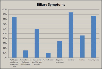
In a study undertaken by Wesdorp et al.4, children with cholelithiasis could be categorized into one of 4 groups based upon their symptoms. 17% remain asymptomatic, 24% will show nonspecific abdominal pain that cannot be defined as colicky, 7% will suffer acute abdominal pain, tenderness and fever, and the remaining 52% will experience biliary symptoms (Fig. 1).
Figure 1: Breakdown of the biliary symptoms identified in children by Wesdorp et al.4 |
|
As you can see from the graph in figure 1, the biliary symptom Wesdorp et al. found to be most prevalent, is jaundice. From this study it is also interesting to note that gallstone frequency increased as the age of the child increased and the female predominance, as seen in adulthood, did not become evident until 14 years of age.
Similarly, Friesen and Roberts14 found that jaundice was the most common symptom of cholelithiasis, but only in infants less than one. They found vomiting to be the most common symptom overall. In a study undertaken by Kumar, Nguyen & Shun15, right upper quadrant pain was found to be the most common symptom, hence illustrating the varied clinical presentation of pediatric cholelithiasis.
The pathophysiological cause of cholelithiasis in a pediatric patient also plays a role in determining the symptoms they may present with. A child may fall into one of three groups depending on the underlying etiology13,15.
- Hemolytic disease: This is considered the most common associated condition, with prevalence rates of 23%15 and 46%14 being reported. Diseases accounted for in this group include sickle-cell disease, thalassemia major, hereditary spherocytosis and rhesus or ABO blood group incompatibility11.
- Specific non-hematological cause: Included in this category are; prematurity, systemic infection, family history, total parenteral nutrition (TPN), pregnancy, oral contraceptive use, obesity, use of the antibiotic ceftriaxone, congenital anomalies of the biliary tract, disease of the terminal ileum such as Crohn’s disease, and surgical resection of the terminal ileum4,11-13,18.
- Idiopathic: Cholelithiasis with no known cause. According to Kumar et al.15 65% of pediatric gallstone cases fall into this category.
If not treated correctly, gallstones may lead to numerous complications. Acute cholecystitis, choledocholithiasis (migration of gallstone(s) into the common bile duct), gallstone pancreatitis12,16 and cholangitis4 (inflammation of the bile ducts) are some of the more common complications. The treatment of choice for symptomatic cholelithiasis, regardless of age, is removal of the gallbladder12,17,18. Known as cholecystectomy, this procedure has been performed on an infant just 16 days old19. This is also the treatment of choice for asymptomatic children when the stones have been present for longer than 12 months16.
Nerve supply of the gallbladder is via three nerves20. Branches of the celiac ganglion supplies sympathetic and visceral afferent fibres, the phrenic nerve supplies somatic afferent fibres, and the vagus nerve is responsible for parasympathetic innervation. Parasympathetic stimulation causes contraction of the gall bladder and an increase in bile secretion.
The vagus nerve is also known as the 10th cranial nerve (CNX) and exits the skull via the jugular foramen between the temporal and occipital bones. Along with supplying parasympathetic control to the gall bladder, CNX also has motor branches to the soft palate and larynx, and sensory fibres to the pharynx and larynx20. Dysfunction of the vagus nerve may thus affect swallowing, speaking, sense of taste, cause hypo-contractility of the gallbladder and reduce bile acid secretion.
Also exiting through the jugular foramen is the accessory nerve (CNXI). Cranial nerve XI controls the upper trapezius and sternocleidomastoid (SCM) muscles along with cervical 1 & 2 nerves (SCM) and cervical 3 & 4 nerves (upper trapezius)21. Both of these muscles control lateral flexion and contralateral rotation of the neck.
During the birth process, a normal procedure that takes place is cranial molding. With each contraction as the infant passes through the birth canal, fluid is forced out of the skull allowing the cranial bones to overlap and reduce the overall cranial size5. If excessive force is not applied to a particular region of the skull, normal size and shape will be achieved within a few days5. However, in the event that the skull is subject to abnormal mechanical forces, caused either internally by the maternal body or from external intervention, cranial alignment may be disrupted. Consequently, cranial nerve entrapment or irritation as it passes through the foramina of the skull may occur7.
Birth interventions such as vacuum and forceps extraction are potential sources of trauma to infants during birth. In a case series of 114 infants presented by Miller et al22, 41% sustained birth intervention. All 114 infants were experiencing suboptimal breastfeeding due to a biomechanical cause, and with a higher than average rate of birth intervention, this group highlight the possible relationship of birth trauma and breastfeeding difficulties.
Although no external force was used during the labor, it is likely that this 7-week-old infant experienced subtle birth trauma. Subtle birth trauma may manifest as mechanical lesions called spinal and cranial subluxations23. Subluxation is when a joint is limited in one or more planes of motion and this fixation has neurologic, vascular, and lymphatic implications on surrounding tissues and organs24. Chiropractors identify and correct spinal and cranial subluxations.
In a study on sub-optimal breastfeeding performed by Vallone24, 18 of the 25 subjects were found to have cervical dysfunction as a result of subluxation of the C1 vertebrae. In 80% of cases improved latch and ability to breastfeed resulted following chiropractic treatment.
Similarly, Holleman et al.25 presented a case where an 8-day-old infant was demonstrating breastfeeding difficulties. The infant was diagnosed with cranial and C1 subluxations. Hewitt23 presented two cases of dysfunctional nursing. One infant was found to have subluxation of the occipital condyles and cranium, and the second infant had C1/2 and cranial subluxations. Likewise, Holtrop26 identified C1/2 and cranial subluxations in an infant with sucking intolerance. In all of these cases, complete resolution of breastfeeding problems resulted from chiropractic treatment. This demonstrates the correlation between upper cervical subluxations, cranial subluxations and breastfeeding dysfunction.
Therefore, taking into consideration the clinical presentation of the 7-week-old infant, it is likely the craniocervical subluxations resulted in a reduced efferent input to the right SCM and/or upper trapezius muscles, limiting their ability to rotate the head to the left. It is acknowledged that a decreased cervical rotation prevents the infant from obtaining a good latch, which is subsequently expressed via the infant having a preference to nurse on a specific breast5, as was the case with this baby girl.
This case demonstrates the importance of infants having full cervical range of motion for optimal breastfeeding. It is proposed that the chiropractic treatment restored proper neuromuscular control of the SCM and upper trapezius muscles thus enabling a painless and complete left rotation of the head and neck.
In theory, it is also plausible to say that correction of cranial misalignments may result in normal functioning of the vagal nerve. Although no direct improvement was identified in the gallbladder function of the infant in discussion, it is proposed that chiropractic treatment may have optimized the capability of the diseased organ. Further research is needed to investigate this concept.
Conclusion
Cholelithiasis is a condition seen in the pediatric population. The age of the child and the underlying pathophysiology impact the clinical presentation and management. In the younger child an asymptomatic presentation and spontaneous resolution is likely. In the older symptomatic child, or in younger children where resolution is not seen after one year, surgical cholecystectomy is the treatment of choice to prevent complications from developing.
Craniocervical subluxations are often identified in infants as a result of the birth process. In this case, chiropractic care restored optimal cervical range of motion and cranial alignment. Consequently, the infant no longer showed a preference to feed on the left breast. This demonstrates how chiropractic treatment may be beneficial in correcting breastfeeding difficulties that have a biomechanical cause.
References:
- Shirmer BD, Winters KL, Edlich R. Cholelithiasis and Cholecystitis. J Long Term Eff Med Implants 2005; 15(3):329-338.
- Koebnick C, Smith N, Black MH, Porter AH, Richie BA, Hudson S, Gililland D, Jacobsen SJ, Longstreth GF. Pediatric obesity and gallstone disease. J Pediatr Gastroenterol Nutr 2012;55(3);328-333.
- Palasciano G, Portincasa P, Vinciguerra V, Velardi A, Tardi S., Baldassarre G, Albano O. Gallstone prevalence and gallbladder volume in children and adolescents; an epidemiological ultrasonographic survey and relationship to body mass index. Am J Gastroenterol 1989;84(11):1378-1382.
- Wesdorp I, Bosman D, de Graaff A, Aronson D, van der Blij F, Taminiau J. Clinical Presentations and Predisposing Factors of Cholelithiasis and Sludge in Children. J Pediatr Gastroenterol Nutr 2000;31:411-417.
- Anrig C, Plaugher G. Pediatric Chiropractic. Philadelphia: Lippincott Williams & Wilkins;2011.
- http://www.who.int/topics/breastfeeding/en/.
- Smith L. Impact of Birthing Practices on the Breastfeeding Dyad. Journal Midwifery & Women’s Health 2007;52;621-630.
- Sacro Occipital Technique Organisation Australasia. S.O.T. Seminar Series Notes. Chiropractic Manipulative Reflex Technique (CMRT) 2009;31-34.
- www.mayoclinic.com/health/hida-scan/MY00320.
- Australian Government Department of Health and Ageing Therapeutic Goods Administration. Australian Public Assessment Report for Ursodeoxycholic acid. 2010.
- Suma V, Marini A, Bucci N, Toffolutti T, Talenti E. Fetal gallstones: sonographic and clinical observations. Ultrasound Obstet Gynecol. 1998;12;439-441.
- Poffenberger CMc, Gausche-Hill M, Ngai S, Myers A, Renslo R. Cholelithiasis and Its Complications in Children and Adolescents. Pediatric Emergency Care 2012;28(1);68-76.
- Poddar U. Gallstone Disease in Children. Indian Pediatrics 2010;47;945-953.
- Friesen CA, Roberts CC. Cholelithiasis. Clinical characteristics in children. Case analysis and literature review. Clin Pediatr (Phila)1989;28(7);294-298.
- Kumar R, Nguyen K, Shun A. Gallstones and common bile duct calculi in infancy and childhood. Aus NZ J Surg 2000;70(3);188-191.
- Bellows CF, Berger DH, Crass RA. Management of Gallstones. American Family Physician. 2005;72(4);637-642.
- Holcomb GW Jr, Holcomb GW 3rd. Cholelithiasis in infants, children and adolescents. Pediatric Rev 1990;11;268-274.
- Della Corte C, Falchetti D, Nebbia G, Calacoci M, Pastore M, Francavilla R, Marcellini M, Vajro P, Iorio R. Management of cholelithiasis in Italian children: A national multicenter study. World J Gastroenterol 2008;14(9);1383-1388.
- Gertner M, Farmer DL. Laparoscopic cholecystectomy in a 16-day-old infant with chronic cholelithiasis. J Pediatr Surg 2004;39(1);E17-19.
- Moore KL, Dalley AF. Clinically Oriented Anatomy (5th ed). Baltimore: Lippincott Williams & Wilkins, 2006: 303.
- Kendall FP, McCreary EK, Provance PG, McIntyre Rodgers M, Romani WA. Muscles Testing and Function with posture and pain. Baltimore: Lippincott Williams & Wilkins, 2005:148-149.
- Miller JE, Miller L, Sulesund A, Yevtushenko A. Contribution of Chiropractic Therapy to Resolving Suboptimal Breastfeeding: A Case Series of 114 Infants. J Manip Physiol Therap 2009;32(8);670-674.
- Hewitt EG. Chiropractic care for infants with Dysfunctional nursing: A case series. J Clin Chiro Paed 1999;4(1);241-244.
- Vallone S. Chiropractic Evaluation and Treatment of Musculoskeletal Dysfunction in Infants Demonstrating Difficulty Breastfeeding. J Clin Chiro Paed 2004;6(1);349-368.
- Holleman AC, Nee J, Knapp SFC. Chiropractic management of breast-feeding difficulties: a case report. J Chiro Med 2011;10;199-203.
- Holtrop DP. Resolution of sucking intolerance in a 6-month-old chiropractic patient. J Manip Physiol Ther 2000;23(9);615-618.
|
